Arterial Supply
Standard hepatic arterial anatomy is present in only about 55–60% of patients. The remainder have anatomical variants that are critically important in transplant surgery, hepatic resection, and embolization procedures.
Standard Anatomy
Celiac trunk → common hepatic artery → proper hepatic artery → right and left hepatic arteries, supplying the corresponding lobes. The gastroduodenal artery (GDA) arises from the common hepatic artery before the bifurcation and serves as an important anatomical landmark.
Replaced Right Hepatic Artery (from SMA)
rRHA — ~15–20%
Click to expand
Replaced Left Hepatic Artery (from LGA)
rLHA — ~12–15%
Click to expand
Accessory Variant Arteries
Supplementary — ~10%
Click to expand
Pre-operative CT angiography (CTA) or MR angiography is standard practice before any major hepatectomy or living donor evaluation specifically to map arterial anatomy.
Portal Venous System
Portal Vein Formation and Flow
Superior mesenteric vein (SMV) + splenic vein → portal vein (formed posterior to the neck of the pancreas)
Portal vein enters the porta hepatis and bifurcates into
right and
left portal veins.
Portal vein provides approximately
75% of hepatic blood flow (high volume, lower oxygen content) — the hepatic artery provides the remaining 25% (lower volume, high oxygen).
Portal pressure measured as
hepatic venous pressure gradient (HVPG) via hepatic vein wedge/free pressure measurement.
HVPG thresholds:- ≥6 mmHg — portal hypertension (abnormal)
- ≥10 mmHg — clinically significant portal hypertension (CSPH); risk of complications escalates
- ≥12 mmHg — threshold associated with variceal formation and bleeding risk; also associated with increased risk of decompensation
Hepatic Veins
Three major hepatic veins drain directly into the inferior vena cava (IVC) at the level of the hepatic confluence, just below the right atrium. There is no portal pedicle structure associated with hepatic veins — they travel within the parenchyma between segments.
The Three Hepatic Veins
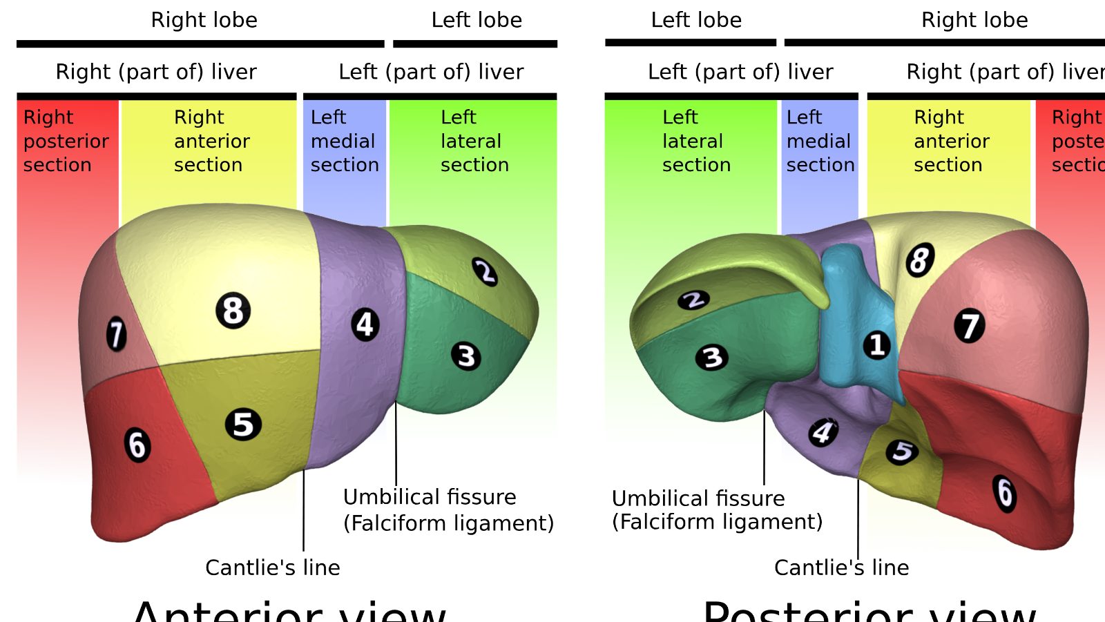
- Right hepatic vein (RHV): Drains segments V, VI, VII, VIII. Largest of the three. Enters the IVC directly on the right. Must be controlled in right hepatectomy.
- Middle hepatic vein (MHV): Drains segments IV, V, and VIII. Courses along the main hepatic scissura (principal plane). Often the key vein in right hepatectomy controversy — whether to include it in the right lobe graft affects outflow and left remnant venous drainage.
- Left hepatic vein (LHV): Drains segments II and III. Frequently joins the middle hepatic vein to form a common trunk before entering the IVC (~80% of individuals).
Surgical Scissurae — Planes of Resection Defined by Hepatic Vein Courses
- Main scissura (principal plane): Runs through the gallbladder fossa to the IVC; courses along the middle hepatic vein. Divides the right and left functional hemilivers. This is the plane of a right or left hepatectomy.
- Right scissura: Separates right anterior (V/VIII) from right posterior (VI/VII) sections; courses along the right hepatic vein.
- Left scissura: Corresponds to the falciform ligament; separates left lateral (II/III) from left medial (IV) sections; courses along the left hepatic vein.
These scissurae are the bloodless planes that surgeons follow during anatomic resections.